Eye Anatomy and Key Diagnoses Rule of 2s
Background
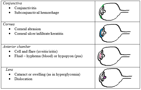
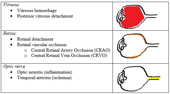
The rule of 2s, as illustrated below, is a simple method with an anatomical approach to the major ocular diagnoses and is meant to provide a general framework to guide the physical exam and include or eliminate certain conditions. The rule of 2s is easy to remember as it contains the number 2 in the context of eyes and proceeds from layer to layer of the eye in an anterior to posterior direction highlighting the 2 major associated diagnoses of that layer.
There is significant overlap among conditions that cause a red, painful or red and painful eye. Here are additional cards that may help in your evaluation:
Anterior Eye – Utilize slit lamp exam
Posterior Eye – Use fundoscopy and ultrasound
References
- Nawar, E.W., Niska, R.W., Xu, J. National Hospital Ambulatory Medical Care Survey: 2005 Emergency Department Summary: Advance Data from Vital and Health Statistics; No. 386. National Center for Health Statistics, Hyattsville, MD; 2007. [PubMed]
- Sharma R, Brunette DD. Ophthalmology. In: Marx JA, Hockberger RS, Walls RM. eds. Rosen's Emergency Medicine - Concepts and Clinical Practice. 8th ed. Philadelphia, PA: Elsevier/Saunders, 2014.
- Walker RA, Adhikari S. Eye Emergencies. In: Tintinalli JE et al, eds. Tintinalli’s Emergency Medicine: A Comprehensive Study Guide. 7th ed. New York: McGraw-Hill, 2011.
