Knee Injuries - Knee Dislocation
Knee Injuries: Knee Dislocation
X-ray Views
AP view
- Look especially for small avulsions, such as a Segond fracture, which is a lateral tibial condyle avulsion fracture
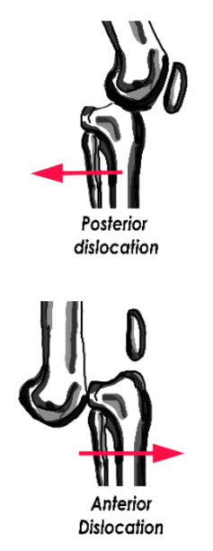
Lateral view
- Look for symmetric or irregular joint space
Acute Management
Orthopedic vascular emergency!
- Reduce knee and re-examine neurovascular status
- Consult vascular surgery for hard signs of vascular injury
Measure the Ankle-Brachial index(ABIs):
- ABI ≥ 0.9: Monitor by serial exam
- ABI < 0.9: Obtain CT angiography
Splint knee in 20-30 degrees of flexion
Follow-Up Timing
Hospital admission required for serial neurovascular exams and consideration for surgical stabilization
Notes
Dimple sign
- Buttonholing of medial femoral condyle through medial capsule
- Suggests irreducible posterolateral dislocation
Associated findings with knee dislocations
- Popliteal artery injury (18%)
- Common peroneal nerve injury (25%) – check for foot dorsiflexion weakness and numbness to dorsal foot
- Ligament tears
References
- Schwartz A. Patella Fractures Treatment & Management, Emedicine.com (June 2016) [Source].
- Hinton RY, Sharma KM. Acute and recurrent patellar instability in the young athlete. Orthop Clin North Am. 2003 Jul;34(3):385-96. [PubMed]
- Ji G, et al . Surgical versus Nonsurgical Treatments of Acute Primary Patellar Dislocation with Special Emphasis on the MPFL Injury Patterns. J Knee Surg. 2016 Sep 14. Epub ahead of print. [PubMed]
- Petri M, et al. Current Concepts for Patellar Dislocation. Arch Trauma Res. 2015 Sep 1;4(3):e29301. [PubMed]
- Medina O, et al. Vascular and nerve injury after knee dislocation: a systematic review. Clin Orthop Relat Res. 2014 Sep;472(9):2621-9. [PubMed]
